
Melbourne Pathology Test Collection Manual: A Comprehensive Plan
Melbourne Pathology’s manual details crucial procedures for accurate testing, encompassing patient preparation, specimen handling, and transport․ It ensures reliable results, adhering to stringent standards for patient safety and diagnostic precision, as of February 27, 2026․
Melbourne Pathology stands as a leading provider of diagnostic pathology services, dedicated to delivering accurate and timely results to healthcare professionals․ This Test Collection Manual serves as a vital resource for all personnel involved in the specimen collection process, ensuring adherence to best practices and maintaining the highest standards of quality․
Our commitment extends beyond simply performing tests; we prioritize patient safety, reliable diagnostics, and collaborative partnerships with clinicians․ This manual reflects that commitment, outlining detailed procedures for every stage – from patient preparation and specimen collection to labeling, transport, and storage․
The information contained herein is based on current guidelines and industry standards, including considerations for radiation safety and the proper handling of various sample types like blood cultures, isolator tubes, and coagulation samples․ We continually update these procedures to incorporate advancements in technology and evolving best practices, referencing publications from organizations like Leica Biosystems and research documented as early as 2019․
This manual is designed to be a comprehensive guide, supporting consistent and accurate testing, ultimately contributing to improved patient care․ It’s essential reading for all phlebotomists, nurses, and other healthcare staff involved in the collection process, ensuring a standardized approach across all locations and departments․
II․ Pre-Analytical Phase: Patient Preparation
The pre-analytical phase is critical for ensuring accurate and reliable test results․ Proper patient preparation directly impacts the quality of specimens collected and, consequently, the diagnostic outcome․ This phase encompasses all procedures before the actual specimen collection, demanding meticulous attention to detail․
Key considerations include verifying patient identity, confirming test requests, and providing clear, concise instructions to patients regarding any necessary preparation․ This often involves specific fasting requirements for certain tests, or advising patients to discontinue specific medications that may interfere with results․
Thorough patient education is paramount․ Patients must understand the purpose of the test, the preparation needed, and any potential risks or discomforts associated with the collection process․ Accurate labeling begins here, with correct patient identification being the foundation of a reliable result․
Failure to adhere to proper pre-analytical procedures can lead to erroneous results, potentially delaying diagnosis or leading to inappropriate treatment․ Melbourne Pathology emphasizes strict adherence to these guidelines, referencing established protocols and continually updating procedures to maintain the highest standards of quality and patient safety, as documented in various publications since 2019․
III․ Patient Identification and Verification
Patient identification is the cornerstone of accurate testing and patient safety within Melbourne Pathology’s framework․ A robust verification process is mandatory before any specimen collection commences․ This involves a multi-faceted approach, utilizing at least two independent identifiers․
Acceptable identifiers include the patient’s full name, date of birth, and a unique medical record number․ Verbal confirmation of these details with the patient is essential, alongside a visual inspection of their identification documentation – such as a driver’s license or healthcare card․ Reliance on patient-reported information alone is insufficient;
Any discrepancies between the information provided by the patient and their identification documents must be resolved before proceeding․ If a reliable identification cannot be established, the collection must be postponed until proper verification is possible․
Accurate labeling, initiated during this phase, is directly linked to correct patient identification․ Melbourne Pathology emphasizes that misidentification can lead to potentially life-threatening errors, highlighting the critical importance of meticulous adherence to these verification protocols, as outlined in their comprehensive collection manual and referenced in publications dating back to 2019․
IV․ Fasting Requirements for Specific Tests
Melbourne Pathology mandates strict adherence to fasting protocols for numerous tests to ensure accurate and reliable results․ Fasting duration varies depending on the specific analysis requested, impacting analyte concentrations and minimizing interference․
Generally, a minimum of 8-12 hours of fasting is required for lipid profiles (cholesterol, triglycerides), glucose measurements, and certain metabolic panels․ During this period, patients are permitted only water intake․ Clear instructions regarding permissible and prohibited substances must be provided to patients before the scheduled collection․
Specific tests, like glucose tolerance tests, necessitate more complex preparation, including timed meals and blood draws․ Failure to comply with fasting instructions can significantly alter test outcomes, leading to misdiagnosis or inappropriate treatment decisions․
The Melbourne Pathology manual details specific fasting requirements for each test, accessible to both patients and healthcare professionals․ It’s crucial to verify these requirements prior to collection․ Documentation of fasting compliance should be recorded alongside the specimen, ensuring traceability and data integrity, as emphasized in their quality control procedures and referenced in materials from 2023․
V․ Medication Interference with Test Results
Melbourne Pathology recognizes that numerous medications can significantly interfere with laboratory test results, potentially leading to inaccurate diagnoses․ Comprehensive patient medication history acquisition is, therefore, paramount during the pre-analytical phase․
Certain drugs can artificially elevate or depress analyte levels, masking underlying conditions or creating false positives/negatives․ For instance, biotin supplementation can interfere with thyroid function tests and cardiac marker assays․ Nonsteroidal anti-inflammatory drugs (NSAIDs) can affect renal function tests and coagulation profiles․
The Melbourne Pathology manual provides a detailed list of medications known to impact common laboratory tests․ Healthcare providers are responsible for assessing potential interference and, when feasible, advising patients to temporarily discontinue medications prior to sample collection – always prioritizing patient safety․
If medication cessation isn’t possible, this information must be meticulously documented on the test requisition form․ This allows the laboratory to interpret results cautiously and potentially adjust testing methodologies․ Accurate medication recording, alongside adherence to collection protocols, is vital for reliable diagnostic outcomes, as highlighted in their 2023 guidelines․
VI․ Specimen Collection Procedures: General Guidelines
Melbourne Pathology emphasizes standardized specimen collection procedures to ensure the integrity and reliability of test results․ All personnel involved in sample acquisition must be thoroughly trained and demonstrate competency in established protocols, as outlined in the comprehensive manual․
General guidelines include maintaining aseptic technique to prevent contamination, utilizing appropriate collection containers with correct preservatives, and adhering to specified volume requirements․ Proper patient positioning and comfort are also crucial for minimizing variables that could influence results․
Detailed instructions are provided for various sample types, including blood, urine, and body fluids․ The manual stresses the importance of minimizing hemolysis during blood collection and avoiding contamination of urine specimens․ All tubes must be labelled immediately after collection with patient identifiers․
Furthermore, adherence to the chain of custody procedures is essential for maintaining sample traceability and legal defensibility․ Melbourne Pathology’s commitment to quality assurance extends to providing ongoing education and updates to collection staff, ensuring consistent and accurate practices, referencing guidelines established as recently as February 27, 2026․
VII․ Blood Collection Techniques
Melbourne Pathology mandates strict adherence to standardized blood collection techniques to guarantee sample quality and patient safety․ Trained phlebotomists must employ proper venipuncture procedures, prioritizing patient comfort and minimizing discomfort throughout the process․
Techniques include careful site selection, typically utilizing the antecubital fossa, and thorough skin disinfection with an appropriate antiseptic solution․ Correct tourniquet application and release are crucial to avoid hemoconcentration․ The manual details proper needle insertion angle and tube handling to prevent contamination or activation of coagulation cascades․
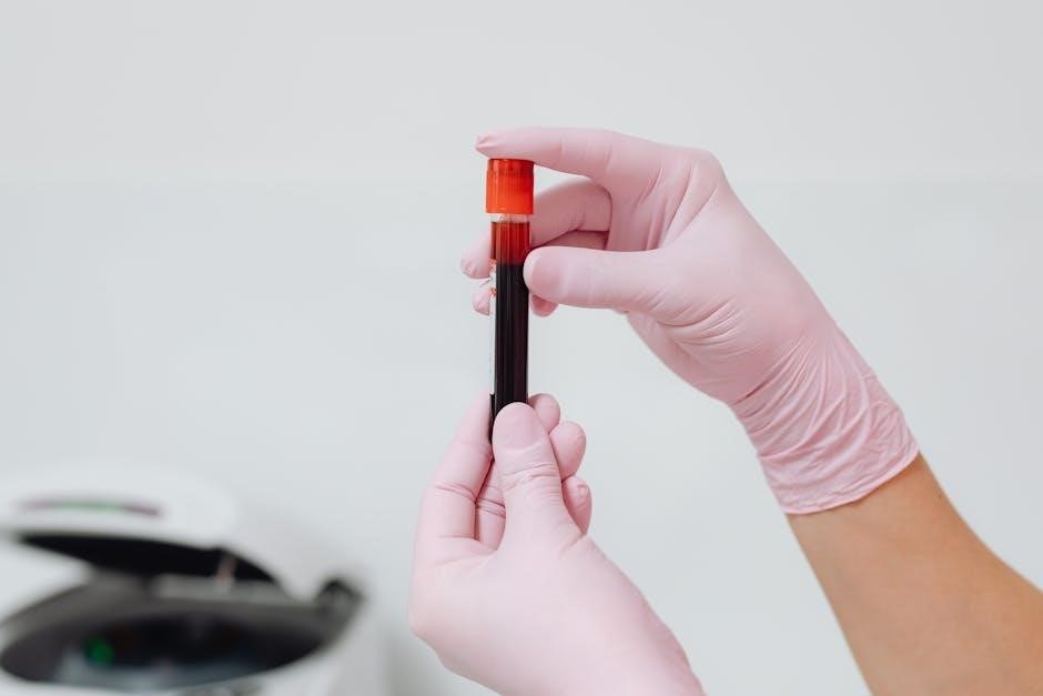
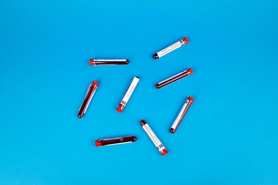
Specific attention is given to avoiding hemolysis, achieved through gentle collection and proper tube mixing․ Following collection, immediate and correct tube inversion is vital for additive-to-sample ratio accuracy․ Blood culture bottles, isolator tubes, and various top tubes (red, gold, blue, royal blue) require distinct handling protocols, as detailed in the manual․
Phlebotomists are expected to recognize and address potential complications, such as hematomas or fainting․ Documentation of any difficulties encountered during collection is essential for accurate interpretation of results, referencing procedures updated as of February 27, 2026․
VIII․ Venipuncture Site Selection
Melbourne Pathology’s guidelines prioritize the antecubital fossa for venipuncture, specifically the median cubital, cephalic, and basilic veins․ The median cubital vein is generally preferred due to its size, stability, and reduced risk of rolling․ Careful assessment of potential sites is crucial, avoiding areas with hematomas, scars, or edema․
Sites on the same side as a mastectomy or lymph node removal should be avoided due to potential lymphedema risk․ If the antecubital fossa is unsuitable, alternative sites like the dorsal hand veins may be considered, but are associated with increased discomfort and potential for hemolysis․
Prior to site selection, a thorough inspection of the arm is required to identify suitable veins․ Palpation should confirm vein bounce and adequate filling․ Avoid veins that are fragile, tortuous, or deeply embedded․ Proper site selection minimizes patient discomfort, reduces the risk of complications, and ensures optimal sample collection․
Documentation of the chosen venipuncture site is mandatory, along with any challenges encountered during site assessment, adhering to protocols updated as of February 27, 2026, for consistent and reliable results․
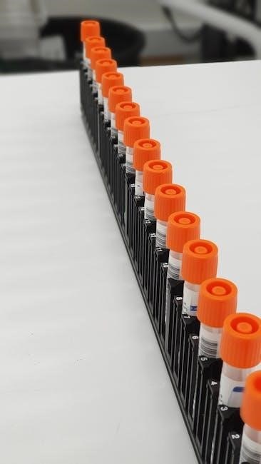
IX․ Order of Draw for Blood Tubes
Melbourne Pathology strictly adheres to a specific order of draw to prevent cross-contamination of additives between blood collection tubes․ The recommended sequence, based on CLSI guidelines, begins with blood culture tubes (yellow-black stopper), followed by coagulation tubes (light blue stopper – sodium citrate)․
Next in sequence are serum tubes (red or gold/SST stoppers), then heparin tubes (green stopper), EDTA tubes (lavender/purple stopper), and finally, glycolytic inhibitor tubes (gray stopper)․ This order minimizes the potential for carryover, ensuring accurate test results․
Proper technique includes gently inverting each tube according to manufacturer’s instructions immediately after collection․ Tubes should be filled to the indicated fill volume to maintain the correct blood-to-additive ratio․ Deviation from the established order of draw can significantly impact test outcomes, particularly coagulation studies․
Detailed adherence to this protocol, updated as of February 27, 2026, is essential for maintaining the highest standards of laboratory accuracy and patient care, as outlined in Melbourne Pathology’s comprehensive manual․
X․ Urine Collection Protocols
Melbourne Pathology emphasizes meticulous urine collection techniques for reliable diagnostic results․ Several protocols exist, tailored to the specific test requested․ The most common is the midstream clean catch, crucial for minimizing contamination from external sources․
Patients are instructed to cleanse the genital area thoroughly before initiating collection, discarding the initial urine stream․ The mid-portion of the subsequent stream is collected into a sterile container․ For 24-hour urine collections, patients receive a container and detailed instructions, including starting time, storage requirements (refrigeration), and completion time․
Complete collection is vital; any missed fractions compromise accuracy․ Timed collections require precise adherence to the specified duration․ Proper labeling with patient identification, date, and time of collection is paramount․ Specific preservatives may be required, as indicated on the test request form․
Adherence to these protocols, updated as of February 27, 2026, ensures the integrity of the sample and the validity of the laboratory findings, as detailed within the Melbourne Pathology manual․
XI․ Midstream Clean Catch Technique
Melbourne Pathology mandates the midstream clean catch technique for most urine analyses to minimize contamination and ensure accurate results․ This method focuses on collecting a sample representative of urine within the bladder, avoiding external influences․
Patients receive clear instructions: begin by washing the genital area thoroughly with soap and water, rinsing completely․ Women should separate labia; men should retract the foreskin․ Initiate urination, allowing a small amount to pass into the toilet․
Immediately after discarding the initial stream, collect the mid-portion of the urine flow into a sterile, provided container․ Avoid touching the inside of the container․ Complete urination after collection․ Securely cap the container and label it accurately with patient details, date, and time․
This technique, crucial for detecting urinary tract infections and other conditions, is detailed in the Melbourne Pathology manual (updated February 27, 2026)․ Proper execution is vital for reliable diagnostic outcomes, minimizing false positives or negatives․
XII․ 24-Hour Urine Collection Instructions
Melbourne Pathology requires precise adherence to 24-hour urine collection protocols for accurate quantitative analyses, such as creatinine clearance and protein quantification․ This involves collecting all urine produced over a full 24-hour period․
Patients begin by emptying their bladder at a specific time, discarding this initial sample․ All subsequent urine, including the first void of the following day at the same time, is collected in a large, provided container, typically containing a preservative․ Maintain refrigeration throughout the collection period to prevent bacterial growth․
Record the start and end times meticulously․ Ensure no urine is lost during collection․ The container must be securely sealed and labeled with patient details, date, and the collection start/end times․
Detailed instructions, updated as of February 27, 2026, are provided by Melbourne Pathology to guarantee reliable results․ Incomplete or improperly collected samples will be rejected, necessitating repeat testing․ Patient compliance is paramount for accurate diagnosis․
XIII․ Specific Sample Types and Handling
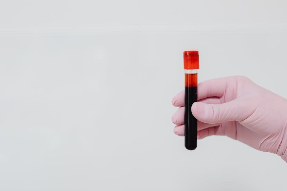
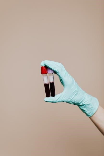
Melbourne Pathology mandates specific handling procedures for diverse sample types to maintain specimen integrity and ensure accurate test results․ Blood culture bottles, isolator tubes, and various blood tubes (blue top – citrate, red top, gold top SST, royal blue top) require distinct protocols․
Blood cultures necessitate aseptic technique during collection to prevent contamination․ Isolator tubes are utilized for microbiology, preserving bacterial viability․ Coagulation studies demand precise filling of blue top (citrate) tubes, maintaining the correct blood-to-anticoagulant ratio․
Serum separator tubes (SST – gold top) require allowing complete clotting before centrifugation to obtain serum․ Red top tubes, lacking preservatives, necessitate prompt processing․ All samples must be labeled correctly, adhering to chain-of-custody procedures․
As of February 27, 2026, Melbourne Pathology emphasizes proper storage temperatures and timely transport to the laboratory․ Detailed guidelines, including those for organ pathology specimens, are available to staff, ensuring optimal sample quality and reliable diagnostic outcomes․
XIV․ Blood Culture Collection & Transport
Melbourne Pathology prioritizes aseptic technique during blood culture collection to minimize contamination, a critical factor for accurate sepsis diagnosis․ Skin antisepsis with chlorhexidine or iodine is mandatory, followed by meticulous venipuncture using sterile equipment․ Multiple blood samples, typically two sets from separate sites, are recommended․
Blood culture bottles must be inoculated promptly after collection, avoiding overfilling or underfilling․ Proper mixing of the sample within the bottle is essential for optimal bacterial growth․ The external surface of the bottles should not be touched after inoculation․
Transport to the laboratory should be immediate․ If a delay is unavoidable, bottles must be maintained at room temperature, avoiding refrigeration or freezing․ As of February 27, 2026, Melbourne Pathology requires clear labeling of blood culture bottles with patient details and collection date/time․

Adherence to these guidelines ensures reliable detection of bloodstream infections, supporting timely and appropriate patient management․ Strict protocols are vital for minimizing false-positive and false-negative results․
XV․ Isolator Tube Usage for Microbiology
Melbourne Pathology utilizes isolator tubes for the collection and transport of specimens intended for microbiological analysis, particularly for fastidious organisms․ These tubes contain a specialized transport medium designed to maintain the viability of microorganisms without allowing overgrowth of contaminating flora․
Proper collection technique is paramount․ The specimen should be introduced into the isolator tube as quickly as possible after collection, avoiding contact with surfaces that may harbor contaminants․ The tube must be tightly sealed to prevent leakage during transport․
Isolator tubes are specifically indicated for specimens such as swabs from wounds, sinuses, or other body sites where a mixed microbial population is anticipated․ They are not suitable for blood cultures, which require dedicated blood culture bottles․
As of February 27, 2026, Melbourne Pathology mandates prompt delivery of isolator tubes to the laboratory․ Refrigeration is generally recommended during transport, but specific storage requirements may vary depending on the organism suspected․ Accurate labeling with patient information and collection details is crucial․
XVI․ Coagulation Sample Collection (Blue Top Tubes)
Melbourne Pathology employs blue top tubes containing 3․2% sodium citrate as the standard for coagulation studies․ This anticoagulant binds calcium, preventing the clotting cascade from proceeding in vitro, thus ensuring accurate test results․
Venipuncture technique is critical․ A gentle draw is essential to avoid tissue trauma, which can activate coagulation factors and lead to falsely elevated results․ A 19- or 21-gauge needle is recommended․ The tube should be filled completely to ensure the correct anticoagulant-to-blood ratio․
Immediately after collection, the blue top tube must be gently inverted 8-10 times to thoroughly mix the blood with the citrate․ Avoid vigorous shaking, which can cause hemolysis․
As of February 27, 2026, Melbourne Pathology requires that coagulation samples be processed within a specified timeframe – typically two hours – to maintain sample integrity․ Delays can affect test accuracy․ Proper labeling and documentation are, as always, paramount for reliable results․
XVII․ Serum Separator Tube (SST) – Gold Top Tubes
Melbourne Pathology utilizes gold top Serum Separator Tubes (SSTs) for a wide range of clinical chemistry and immunological assays․ These tubes contain a gel separator that automatically separates serum from cells after centrifugation, simplifying the process and minimizing contamination․
Proper filling is crucial; underfilled tubes can alter the serum-to-gel ratio, impacting test results․ A gentle draw during venipuncture is recommended to avoid hemolysis, which can interfere with many assays․ As of February 27, 2026, Melbourne Pathology mandates a specific order of draw to prevent cross-contamination of additives from other tubes․
Following collection, allow the blood to clot completely – typically 30 minutes – before centrifugation․ Centrifuge at the speed and duration specified in the Melbourne Pathology guidelines․ Avoid disturbing the tube during centrifugation to prevent mixing of serum and cells․
Serum should be separated from the gel and transferred to a clean container for analysis․ Accurate labeling and meticulous documentation remain essential for ensuring the integrity and traceability of each sample․
XVIII․ Sodium Citrate Tubes (Blue Top) – Coagulation Studies
Melbourne Pathology employs blue top Sodium Citrate tubes specifically for coagulation testing, including Prothrombin Time (PT), Activated Partial Thromboplastin Time (aPTT), and fibrinogen levels․ The citrate acts as an anticoagulant by binding calcium, essential for the coagulation cascade․
Maintaining a precise blood-to-anticoagulant ratio is paramount; deviations can lead to inaccurate results․ These tubes require a full draw to ensure the correct ratio․ Gentle mixing immediately after collection is vital to prevent clot formation, but vigorous shaking should be avoided to prevent platelet activation․
As per Melbourne Pathology’s protocol, dated February 27, 2026, samples must be tested within a defined timeframe after collection to maintain accuracy․ Prolonged storage can affect coagulation factor activity․ Proper centrifugation is also crucial, following established guidelines․
Careful handling and adherence to the specified order of draw are essential to prevent contamination from other tube additives․ Accurate labeling and complete documentation are non-negotiable for reliable coagulation test results․
XIX․ Specimen Labeling and Documentation
Melbourne Pathology emphasizes meticulous specimen labeling and documentation as foundational to accurate diagnostic results․ Every sample, collected as of February 27, 2026, must be unequivocally identified with at least two unique identifiers – typically the patient’s full name and a unique medical record number․
Labels must be affixed directly to the specimen container, ensuring they are securely attached and legible․ Handwritten labels are permissible only when pre-printed labels are unavailable, requiring clear and concise handwriting․ Any discrepancies between the label and the request form necessitate immediate resolution․
Complete documentation, including the date and time of collection, collector’s initials, and any relevant clinical information, is mandatory․ This information is crucial for interpreting test results and maintaining a robust audit trail․
Furthermore, Melbourne Pathology adheres to strict chain of custody procedures, particularly for forensic or legal specimens, ensuring sample integrity from collection to analysis․ Proper documentation safeguards patient safety and supports legally defensible results․
XX․ Importance of Accurate Labeling
Melbourne Pathology underscores that accurate specimen labeling is paramount, directly impacting patient safety and the reliability of diagnostic outcomes, as of February 27, 2026․ Mislabeling can lead to incorrect test results, potentially causing misdiagnosis, inappropriate treatment, and significant patient harm․
A correctly labeled specimen ensures the laboratory can confidently link the sample to the correct patient record and clinical request․ This minimizes the risk of errors throughout the testing process․ The manual stresses the necessity of verifying label information against the patient’s identification band and the test requisition form before sample collection․
Incomplete or illegible labels are unacceptable and will result in sample rejection․ This prevents delays in diagnosis and avoids unnecessary repeat collections․ Consistent adherence to labeling protocols demonstrates a commitment to quality assurance and patient care․
Ultimately, meticulous labeling is not merely a procedural step; it’s a critical component of responsible laboratory practice, upholding Melbourne Pathology’s dedication to accurate and dependable results․
XXI․ Chain of Custody Procedures
Melbourne Pathology implements rigorous chain of custody procedures to maintain specimen integrity and ensure legally defensible test results, current as of February 27, 2026․ This protocol meticulously documents the handling of each sample, from collection to analysis, establishing an unbroken trail of possession․
The chain of custody begins with the collector, who is responsible for initial labeling and documentation․ Each transfer of the specimen – to transport, receiving, or the laboratory – must be recorded with the date, time, and signatures of both the relinquishing and receiving personnel․
Any deviation from the standard protocol, such as a break in the documented chain, compromises the specimen’s admissibility for legal purposes․ Secure packaging and temperature control during transport are also integral components of maintaining the chain of custody․
Melbourne Pathology’s commitment to these procedures guarantees the authenticity and reliability of test results, particularly crucial in forensic, legal, and regulatory contexts․ Strict adherence safeguards against tampering and ensures accountability at every stage․
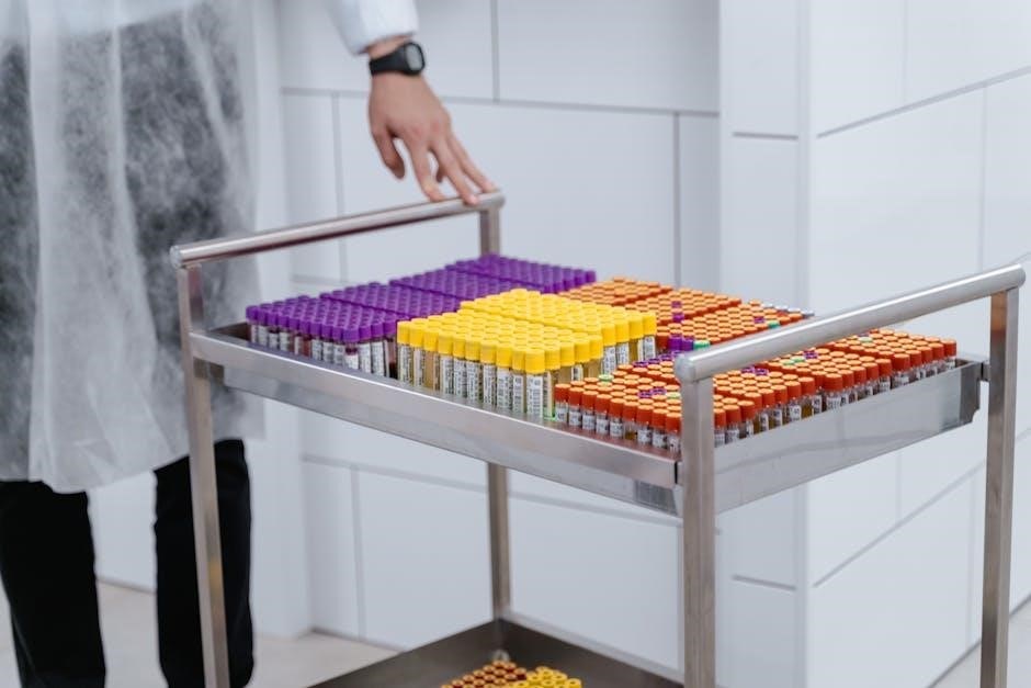
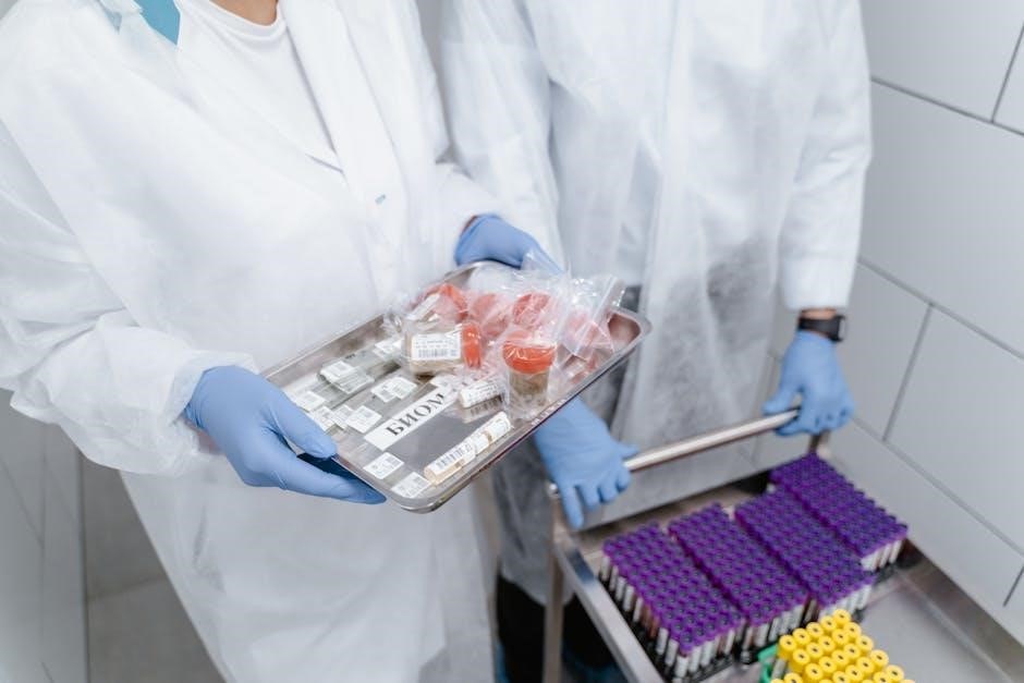
XXII․ Post-Analytical Phase: Specimen Transport & Storage
Melbourne Pathology’s post-analytical phase, as of February 27, 2026, focuses on maintaining specimen integrity during transport and storage, crucial for accurate results․ Proper handling post-collection is paramount to prevent degradation or contamination, impacting diagnostic reliability․
Specimen transport adheres to strict temperature guidelines, utilizing validated containers and transport media․ Refrigerated or frozen samples require continuous temperature monitoring, documented throughout the journey․ Timely delivery to the laboratory is essential, minimizing delays that could affect sample viability․
Upon arrival, specimens are logged, and storage conditions are meticulously maintained․ Storage durations vary based on the test type and sample matrix, following established protocols․ Accurate inventory management and controlled access to storage areas are vital․
Melbourne Pathology employs validated storage systems and regularly monitors equipment to ensure optimal conditions․ Detailed records of storage locations, dates, and temperatures are maintained, guaranteeing traceability and compliance with regulatory standards․ This phase ensures the integrity of the sample until analysis․